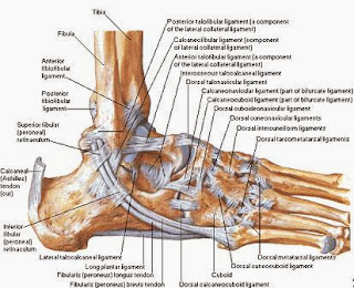
Medial ankle ligaments
The medial ankle ligaments (or deltoid ligament) is a strong, flat, triangular band, attached, above, to the apex and anterior and posterior borders of the medial malleolus. The deltoid ligament is composed of the anterior tibiotalar ligament, tibiocalcaneal ligament, posterior tibiotalar ligament and the tibionavicular ligament. It consists of two sets of fibres, superficial and deep.Important ligaments of the ankle are those that compose the distal tibiofibular syndesmosis and the lateral and medial collateral ligaments (1,2). The lateral collateral ligament consists of three ligaments: the anterior talofibular ligament, the posterior talofibular ligament, and the calcaneofibular ligament. The medial collateral ligament (deltoid ligament) consists of four ligaments: the anterior tibiotalar ligament, the posterior tibiotalar ligament, the tibionavicular ligament, and the tibiocalcaneal ligament. Assessment of the extent of ligamentous injury has been accomplished with routine radiography, stress radiography, arthrography, and tenography (3,4). Owing to the advantage of detailed demonstration of soft-tissue structures and the capability for direct multiplanar demonstration of the medial ankle ligaments, magnetic resonance (MR) imaging has been increasingly applied to the evaluation of ligamentous injuries of the ankle. The ankle joint consists of the tibia and fibula articulating with the talus, also known as the talocrural joint. This joint has many ligaments and a surrounding thick capsule to provide stability and absorb the high amounts of force traveling thought the joint.
The Primary Medial Ankle Ligaments
The primary medial ankle ligaments is the deltoid ligament, which is comprised of the anterior and posterior tibiotalar ligaments, the tibiocalcaneal ligament, and the tibionavicular ligaments. These ligaments branch from medial malleolus of the tibia to the talus, calcaneus, and navicular. Cadaver studies have shown up to 4 superficial and 2 deep bands can be found. The superficial ligaments cross the talocrural and subtalar joint, whereas the deep ligaments only cross the subtalar joint. Hintermann et al. identifies the 6 ligamentous layers: tibionavicular, tibiospring, tibiocalcaneal, anterior tibiotalar, superficial tibiotalar, and posterior tibiotalar. Between all these fibers the deltoid ligament limits the ankle’s eversion as well as plantarflexion and dorsiflexion.The Incidence
The ankle is one of the most commonly injured joints with sprains accounting for 77% of all ankle injuries5 and for 10-30% of all injuries in sport:1,6,7,22. 10-18% of ankle sprains are medial sprains and are typically associated with subsequent syndesmosis injury or medial maleolus fracture. 85% of ankle sprains are lateral sprains. Specific populations and sports display an increased incidence of medial ankle sprains:
male gender
higher competition levels
young populations
intercollegiate rugby
Acute Ankle Injury
Acute injury can be caused by mechanical forces that exceed the ligamentous, capsular, and bony strength. This injury can occur during2
Competitive sports that require jumping and cutting activities.
Landing on uneven surfaces
Misstep on stairs
Chronic Ankle Injury
Chronic ankle instability/dysfunction is often present after ligamentous injury. It has been reported that there is a 50% incidence of chronic ankle dysfunction after injury to the medial ankle ligaments. Research shows that functional instability is present in 29-42% of people with previous sprained ankles.8 Mechanical factors can include valgus of the foot, and medial ankle laxity.9 Symptoms include:Pain with activity
Recurrent swelling
Complaints of "giving way"
Repetitive injuries

